АвтоАвтоматизацияАрхитектураАстрономияАудитБиологияБухгалтерияВоенное делоГенетикаГеографияГеологияГосударствоДомДругоеЖурналистика и СМИИзобретательствоИностранные языкиИнформатикаИскусствоИсторияКомпьютерыКулинарияКультураЛексикологияЛитератураЛогикаМаркетингМатематикаМашиностроениеМедицинаМенеджментМеталлы и СваркаМеханикаМузыкаНаселениеОбразованиеОхрана безопасности жизниОхрана ТрудаПедагогикаПолитикаПравоПриборостроениеПрограммированиеПроизводствоПромышленностьПсихологияРадиоРегилияСвязьСоциологияСпортСтандартизацияСтроительствоТехнологииТорговляТуризмФизикаФизиологияФилософияФинансыХимияХозяйствоЦеннообразованиеЧерчениеЭкологияЭконометрикаЭкономикаЭлектроникаЮриспунденкция
Method of research in a view of a luminescence
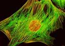 The method of research in a view of a luminescence (luminescent microscopy, or fluorescent microscopy) consists in supervision under a microscope of a green-orange luminescence of microobjects which arises at their illumination by blue-violet light or not visible an eye ultra-violet beams. Two optical filters are entered into the optical scheme of a microscope. One of them place before condenser. It passes from a source-gaffer radiation only those lengths of waves which raise a luminescence of the object (own luminescence), or the special dyes entered into a preparation and absorbed by its particles (secondary luminescence). The second optical filter which is established after an objective, passes to an eye of the observer (or on a photosensitive layer) only light of a luminescence. In luminescent microscopy illumination of preparations from above (through an objective which in this case serves and condenser), and from below, through usual condenser are used. Supervision at illumination from above sometimes name «luminescent microscopy in reflected light» It often share with supervision on a phase-contrast method in passing light. The method is widely applied in microbiology, virology, histology, cytology, in the food-processing industry, in the microchemical analysis, in defectoscopy. Such variety of applications speaks very high color sensitivity of an eye and high picture contrast of self-shone object a dark not luminescing background. Besides the information on structure and properties of investigated substances which can be received, knowing intensity and spectral structure of their luminescent radiation, has huge value.
Поиск по сайту: